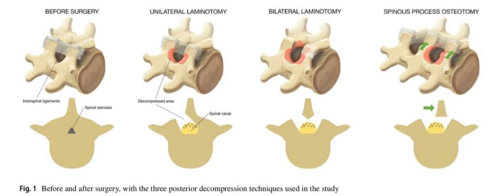
Aumentos comparables en el área del saco dural después de tres técnicas diferentes de descompresión posterior para la estenosis espinal lumbar: resultados radiológicos de un ensayo aleatorio controlado en el estudio NORDSTEN
Investigar los cambios en el área del saco dural después de tres técnicas diferentes de descompresión posterior en pacientes sometidos a cirugía por estenosis espinal lumbar. La descompresión de las raíces nerviosas es el principal tratamiento quirúrgico para la estenosis espinal lumbar. El objetivo de este estudio fue investigar radiológicamente tres técnicas de descompresión posterior comúnmente utilizadas.
Para los pacientes con estenosis espinal lumbar, las tres técnicas quirúrgicas diferentes proporcionaron el mismo aumento en el área del saco dural.
https://pubmed.ncbi.nlm.nih.gov/32556585/
https://link.springer.com/article/10.1007%2Fs00586-020-06499-0
Hermansen E, Austevoll IM, Hellum C, et al. Comparable increases in dural sac area after three different posterior decompression techniques for lumbar spinal stenosis: radiological results from a randomized controlled trial in the NORDSTEN study [published online ahead of print, 2020 Jun 18]. Eur Spine J. 2020;10.1007/s00586-020-06499-0. doi:10.1007/s00586-020-06499-0
Open Access This article is licensed under a Creative Commons Attribution 4.0 International License, which permits use, sharing, adaptation, distribution and reproduction in any medium or format, as long as you give appropriate credit to the original author(s) and the source, provide a link to the Creative Commons licence, and indicate if changes were made. The images or other third party material in this article are included in the article’s Creative Commons licence, unless indicated otherwise in a credit line to the material. If material is not included in the article’s Creative Commons licence and your intended use is not permitted by statutory regulation or exceeds the permitted use, you will need to obtain permission directly from the copyright holder. To view a copy of this licence, visit http://creativecommons.org/licenses/by/4.0/.
Copyright © 2020, Springer Nature


 Dedicado de tiempo completo a atender los problemas de la Columna Vertebral en Adultos y Niños, es decir Diagnóstico, Prevención y Tratamiento de las Enfermedades de la Columna Vertebral en Adultos y Niños. Ejerciendo este quehacer desde hace 25 años.